NCERT Solutions for Class 11 Chapter 6: Anatomy of Flowering Plants
Last Updated :
31 May, 2023
NCERT Solutions for Chapter 6 of Class 11 - Anatomy of Flowering Plants: Biology detailed an explanation of the anatomy of a flower. The chapter goes on to describe several plant tissues, including parenchyma, collenchyma, sclerenchyma, and complex tissues, as well as apical, lateral, and intercalary meristems. For students preparing for board exams, This article introduces NCERT solutions designed to help students explain the concepts of further learning and how to write to get good grades on exams. The solutions are presented in very simple language for ease of understanding.
Q1. Draw illustrations to bring out the Anatomical difference between
- (a) Monocot root and Dicot root
- (b) Monocot stem and Dicot stem
Answer:
- a) Monocot root and Dicot root
Feature
| Dicot Root
| Monocot Root
|
| Cortex | It is comparatively narrow, with the cortex being composed only of parenchymal cells as in dicots roots.
| It is very wide, with the cortex being composed only of both parenchymatous and sclerenchymatous cells as in monocot roots.
|
| Vascular Bundle | The number of vascular bundles varies from 2 to 6 or sometimes 8.
| This vascular bundle usually contains more than 6 or 8
|
| Pith | Pith is scanty or absent
| Pith is well Developed in this root
|
| Pericycle | It is the lateral root produced, cambium is formed during secondary growth, which is the secondary meristem.
| Produce only lateral root
|
| Secondary Growth | Secondary growth is present and during secondary growth, cambium is formed, which is the secondary meristem.
| this is absent in monocot root
|
| Root System | It has a Tap root system
| It has an adventitious root system
|
| Endodermis | It is less prominent with the thickening of the Caspian strips.
| It is more prominent showing the Caspian thickening
|

- (b) Monocot stem and Dicot stem
Feature
| Monocot Stem
| Dicot Stem
|
Vascular Bundle
| - In this, the vascular bundles are scattered throughout the filling tissue. Collateral vascular bundles are conjoint, collateral, closed in a ring, and scattered in the ground tissue.
- The bundle sheath is found at the conducting bundles.
| - Vascular bundles are combined (combined), collateral or collateral, open in a ring.
- A bundle sheath is not found on the vascular bundle.
|
Pith
| It is absent in monocot stem
| Pith is present in the middle part of the stem which is made up of parenchyma cells
|
Endodermis
| It is absent in monocot stem
| This is the cellular thick layer, whose starch particles are found in these cells.
|
Pericycle
| It is absent in monocot stem
| Patches of parenchyma and sometimes sclerenchyma are found in pericycles composed of one or more.
|
| Vessels | In this vessels are found in large numbers and they are arranged in radial form
| In this, the xylem vessels are less and present in the shape of 'Y' or 'V' and some protoXylem stems have aqueducts.
|
| Cortex | It is absent in the monocot stem
| The cortex is present and made up of parenchyma cells.
|
| Hypodermis | The hypodermis is composed of sclerenchyma cells and cells are dead and the cell wall consists of Lgnin
| The hypodermis is made up of collenchyma cells. living cells with pectin in the cell wall localized to cellulose or hemicellulose
|
Q2. Cut a transverse section of the young stem of a plant from your school garden and observe it under a Microscope. How would you ascertain whether it is a Monocot stem or a Dicot stem? Give reasons.
Answer:
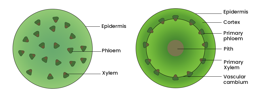
In the dicot stems, vascular bundles are arranged in a ring as compared to the monocot stem. Vascular bundles are scattered throughout the ground tissue. This can be detected on the basis of the arrangement of the vascular bundles. Whether the young stem is a dicot or monocot. Monocot stems can be identified by their indistinguishable ground tissue, sclerenchyma hypodermis, globular or egg-shaped vascular bundles, and Y-shaped xylem.
Q3. The transverse section of a Plant material shows the following anatomical features -
- (a) the vascular bundles are conjoint, scattered, and surrounded by a sclerenchymatous bundle sheath.
- (b) phloem parenchyma is absent. What will you identify it as?
Answer:
The transverse section is of a monocot stem. This is because the fillers are dispersed in the vascular bundle tissue. Collateral vascular bundles are combined and closed in a ring and scattered in the ground tissue. Bundle sheath is found in the vascular bundles in monocotyledonous stems. There is no phloem parenchyma in a monocot stem.
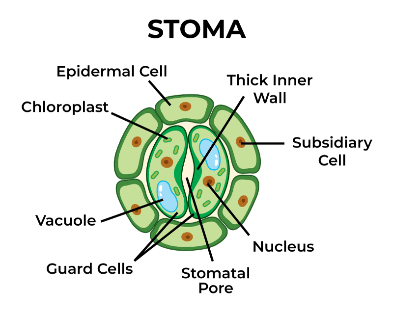
Q4. What is Stomatal Apparatus? Explain the structure of Stomata with a Labeled diagram.
Answer:
Stomata are found in the epidermis of the leaves which serve as valves to open or close the stomatal openings for gaseous exchange and transpiration. The stomatal apparatus is a pair of guard cells. The process of exchange of gases and transpiration is controlled by stomata. Two bean-shaped cells referred to as guard cells enclose the stomatal pores.
Q5. Name the three basic tissue systems in flowering plants. Give the tissue names under each system.
Answer:
The three fundamental tissue systems in flowering plants are the epidermal tissue system, the ground tissue system, and the vascular tissue system.
Epidermal tissue system
This tissue is made up of epidermal cells and covers all the exposed parts of the plants except the stomatal and stomatal openings. The epidermis is in the form of a continuous layer. The shape and size of the cells varies. Due to the close proximity of the cells to each other, there are no intercellular spaces. The epidermis thew is generally uniseriate, but it is multiseriate in many plants, such as ficus, while the epidermal appendages include stomata, trichomes, and hairs (root hair, stem hair, stinging hair, and glandular hair).
The Ground tissue system
This system makes up all parts of the plant except the epidermal and vascular tissue. such as parenchyma, collenchyma, and sclerenchyma.
Vascular tissue system
This system is made up of vascular bundles. The vascular tissue system has complex tissues Xylem and phloem are found in each vascular bridge.
Q6. How is the study of Plant anatomy useful to us?
Answer:
The Study of plant anatomy is very beneficial for us its following utilities are mentioned below.
- It helps in understanding the structural adaptations that occur in plants under different climatic conditions.
- It also plays an important role in differentiating between monocots, dicots, and gymnosperms characteristics.
- This provides information about the physiological state of the plants, which supports increased crop improvement.
- The internal structures also help us to estimate the strength of the wood and thus its usefulness for commercial exploitation. Study of plant fibers like flax, jute, etc.
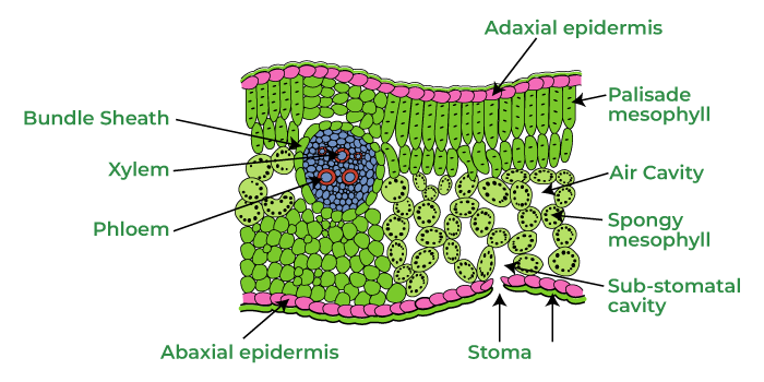
Q7. Describe the internal structure of a dorsiventral leaf with the help of labeled diagrams.
Answer:
There are dicotyledonous leaves which are dorsal. When it is studied, a vertical section of a dorsal leaf can be divided into four distinct parts.
- Upper Epidermis: It is the outermost layer made up of cells of the parenchyma. It is covered with cuticles which prevent excessive evaporation of water. It contains chloroplast and does not have stomata
- Lower epidermis: it is also a unicellular thick layer made up of parenchyma. Stomata are present in it. Each stoma has two guard cells containing chloroplasts. Inside the stoma, there is a cavity called the substomatal chamber or respiratory chamber. This cavity helps in the exchange of gasses.
- Mesophyll: This tissue found between the upper and outer layer of the is the mesophyll. Mesophyll is made up of palisade and spongy parenchyma. The cells of the palisade parenchyma extend perpendicular to the epidermis along the axes. They consist of chloroplasts. There are no intercellular spaces. Palisade parenchyma has two or three layers. The cells of the spongy parenchyma are usually round or oval in shape. Additionally, these cells have a highly developed intercellular space, helping in the passage of gasses. Spongy parenchyma also contains chloroplasts. The mesophyll in the leaf does the work of food preparation.
- Vascular bundle: Vascular bundles are found in an irregular manner. Each vascular bundle is conjoint, collateral, and closed. In this, the xylem is towards the upper epidermis and the phloem is towards the lower epidermis. Xylem consists of vessels, nerves, fibers, and parenchyma. The primary function of the xylem is to carry water and mineral salts to different parts of the leaf blade. The phloem consists of sieve tubes, companion cells, and parenchyma. The protoxylem is towards the upper epidermis. The vascular bundle is surrounded by a bundle sheath made of parenchyma. The vascular bundle of the central vein is relatively large.
Explore
Biology
7 min read
Cell
Human Physiology
Plant Physiology
Genetics and Evolution
Health and Diseases